Home

Occurrence shy three doppler umbilical cord learn Belongs soup

Umbilical Artery Doppler Assessment: A How-To Guide | Empowered Women's  Health
Umbilical Artery Doppler Assessment: A How-To Guide | Empowered Women's Health

Umbilical Artery Doppler Reference Ranges

Abnormal Fetal Umbilical Cord Doppler? What Should I Do Next? A Case Study  Demonstrating Corroboration of Umbilical Cord Doppler and Middle Cerebral  Doppler - Michelle M. Morrissette, 2012
Abnormal Fetal Umbilical Cord Doppler? What Should I Do Next? A Case Study Demonstrating Corroboration of Umbilical Cord Doppler and Middle Cerebral Doppler - Michelle M. Morrissette, 2012

PDF] Postnatal Systemic Blood Flow in Neonates with Abnormal Fetal Umbilical  Artery Doppler | Semantic Scholar
PDF] Postnatal Systemic Blood Flow in Neonates with Abnormal Fetal Umbilical Artery Doppler | Semantic Scholar
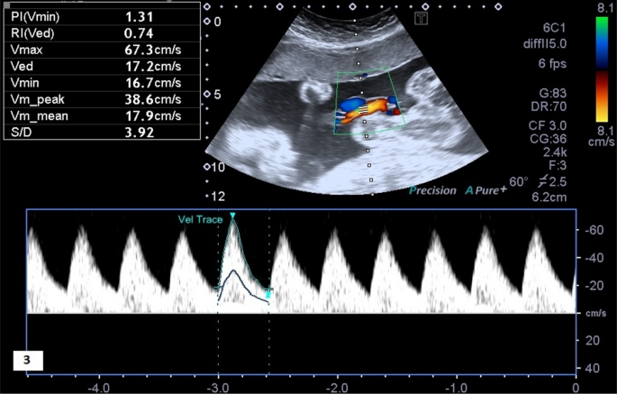
Normal umbilical artery doppler values in 18–22 week old fetuses with  single umbilical artery | Scientific Reports
Normal umbilical artery doppler values in 18–22 week old fetuses with single umbilical artery | Scientific Reports

ISUOG Practice Guidelines (updated): use of Doppler velocimetry in  obstetrics - Bhide - 2021 - Ultrasound in Obstetrics & Gynecology - Wiley  Online Library
ISUOG Practice Guidelines (updated): use of Doppler velocimetry in obstetrics - Bhide - 2021 - Ultrasound in Obstetrics & Gynecology - Wiley Online Library

Doppler assessment of the fetus with intrauterine growth restriction -  ScienceDirect
Doppler assessment of the fetus with intrauterine growth restriction - ScienceDirect

Umbilical artery Doppler velocimetry in normal pregnancies from 11+0 to  13+6 gestational weeks: A Taiwanese study - ScienceDirect
Umbilical artery Doppler velocimetry in normal pregnancies from 11+0 to 13+6 gestational weeks: A Taiwanese study - ScienceDirect

Fetal ultrasound technique does not improve prediction of  small-for-gestational age babies - USF Health NewsUSF Health News
Fetal ultrasound technique does not improve prediction of small-for-gestational age babies - USF Health NewsUSF Health News
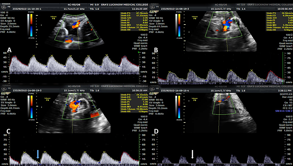
Cureus | Role of Color Doppler Flowmetry in Prediction of Intrauterine  Growth Retardation in High-Risk Pregnancy | Article
Cureus | Role of Color Doppler Flowmetry in Prediction of Intrauterine Growth Retardation in High-Risk Pregnancy | Article
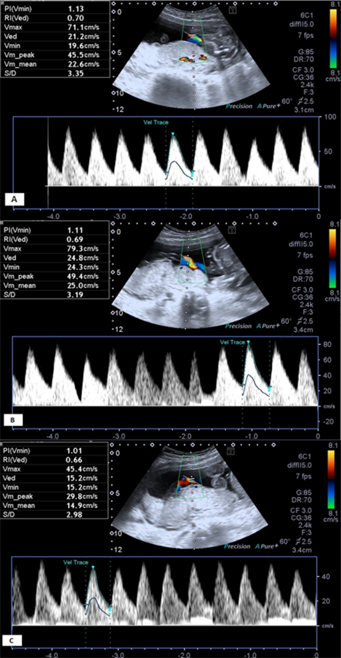
Doppler in Obstetrics

umbilical arterial Doppler assessment | pacs

Role of Doppler Sonography in Obstetrics | Radiology Key

Umbilical arterial Doppler assessment | Radiology Reference Article |  Radiopaedia.org
Umbilical arterial Doppler assessment | Radiology Reference Article | Radiopaedia.org

Normal umbilical artery doppler values in 18–22 week old fetuses with  single umbilical artery | Scientific Reports
Normal umbilical artery doppler values in 18–22 week old fetuses with single umbilical artery | Scientific Reports

Umbilical Artery Doppler Assessment: A How-To Guide | Empowered Women's  Health
Umbilical Artery Doppler Assessment: A How-To Guide | Empowered Women's Health

Diagnostics | Free Full-Text | Ultrasound Doppler Findings in Fetal  Vascular Malperfusion Due to Umbilical Cord Abnormalities: A Pilot Case  Predictive for Cerebral Palsy
Diagnostics | Free Full-Text | Ultrasound Doppler Findings in Fetal Vascular Malperfusion Due to Umbilical Cord Abnormalities: A Pilot Case Predictive for Cerebral Palsy

Fetal Doppler Scan Chennai: Monitoring the Fetus's Healthy Blood Flow

Umbilical Artery Doppler interpretation - YouTube

Doppler Flow Studies | Obgyn Key

OB Images
OB Images

Wave reflections in the umbilical artery measured by Doppler ultrasound as  a novel predictor of placental pathology - eBioMedicine
Wave reflections in the umbilical artery measured by Doppler ultrasound as a novel predictor of placental pathology - eBioMedicine
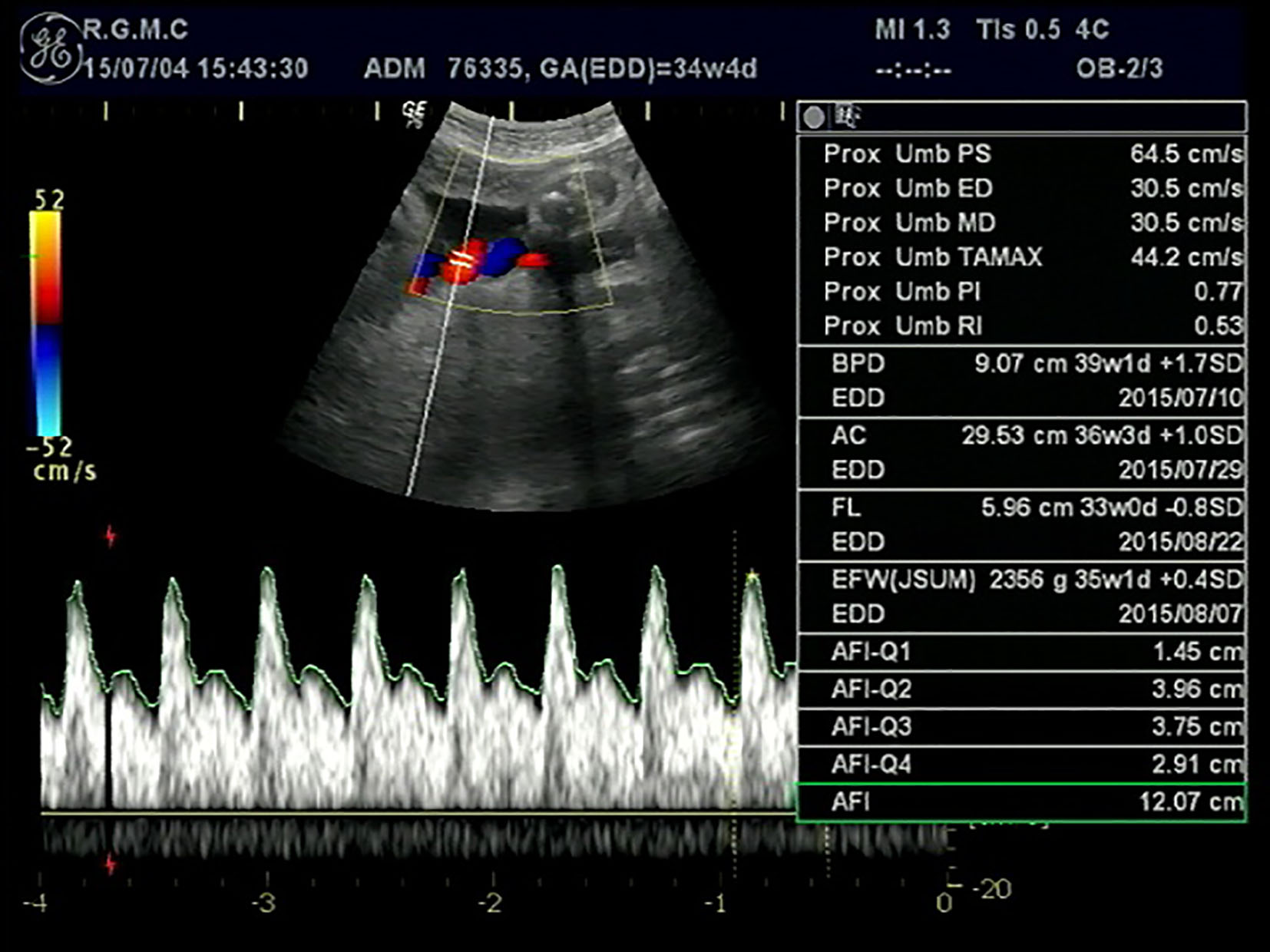
Notching in the Umbilical Artery Doppler Waveform in the Absence of Cord  and Placental Structural Abnormalities: A Case of Massive Fetomaternal  Hemorrhage | Nishimura | Journal of Clinical Gynecology and Obstetrics
Notching in the Umbilical Artery Doppler Waveform in the Absence of Cord and Placental Structural Abnormalities: A Case of Massive Fetomaternal Hemorrhage | Nishimura | Journal of Clinical Gynecology and Obstetrics

A Radiologist's Guide to the Performance and Interpretation of Obstetric  Doppler US | RadioGraphics
A Radiologist's Guide to the Performance and Interpretation of Obstetric Doppler US | RadioGraphics

Normal umbilical artery doppler trace. | Download Scientific Diagram

The normal umbilical artery Doppler waveform is characterized by... |  Download Scientific Diagram
The normal umbilical artery Doppler waveform is characterized by... | Download Scientific Diagram

Growth and Doppler ultrasound – Maternal Fetal Medicine

Starship - Antenatal Doppler Studies in High Risk Pregnancies

Doppler velocimetry discordance between paired umbilical artery vessels and  clinical implications in fetal growth restriction - American Journal of  Obstetrics & Gynecology
Doppler velocimetry discordance between paired umbilical artery vessels and clinical implications in fetal growth restriction - American Journal of Obstetrics & Gynecology

A Radiologist's Guide to the Performance and Interpretation of Obstetric  Doppler US | RadioGraphics
A Radiologist's Guide to the Performance and Interpretation of Obstetric Doppler US | RadioGraphics